


Osteoarthritis of the hip joint (ATS) is a progressive degenerative disease. Under the influence of several reasons, during the development of the disease, irreversible changes in the structure and properties of hyaline cartilage occur, which leads to increased pressure on the articular surface and its deformation or fusion. Since mechanical load is considered one of the main causes of the development of the disease, the articulation of the hip joint is often affected by arthrosis.
Features of the anatomical structure of the hip joint
The hip joint (TC) is the junction of the pelvis and femur. This articulation makes it possible to reduce and spread the lower limbs, lift the legs and pull them to the body, and make walking movements. From birth and throughout life, a person bears a high load on the hip joint.
From the side of the pelvic bone, the "acetabular" cavity takes part in the articulation, from the side of the femur, its epiphysis. The acetabulum has a lip of collagen along the edge, which acts as a kind of gasket that tightly holds the epiphysis of the femur in its recess. The recess in the middle of the acetabulum is covered with a collagen membrane and is the site of attachment of the femoral ligament.
The composition of the TS capsule includes ligaments:
- femoral-iliac - the strongest ligament that can withstand a load of more than 200 kg and prevent excessive backward bending of the hip;
- femoral-pubic - responsible for abduction and reduction of the thigh, thereby limiting its circular movement;
- femoral ischial - protects the vehicle from vibrations, reduces the load when walking and running;
- round (loop) - prevents dislocation and holds the head of the femur in the cavity of the pelvic cavity and is the basis of the articular bag.
Many groups of muscles and tendons allow the vehicle to move around three axes:
- Longitudinal (vertical).
- Transverse (horizontal, forward).
- Sagittal (anterior-posterior).
Joint arthrosis can occur both in healthy joints and be a continuation of existing diseases of the musculoskeletal system.
What is this disease?
Hyaline cartilage performs the function of shock absorption and protection against damage to the joint surface. ATS is a disease in the developmental process in which the structure of collagen cartilage fibers changes, which then leads to their breakdown and destruction. Fragments of cartilage fibers, if they enter the articular cavity, can cause an inflammatory process. The bare surface undergoes changes in bone tissue caused by friction and increased pressure. The remaining cartilaginous tissue along the edge of the epiphysis grows compensatory with subsequent hardening, causing ankylosis (immobility of the bone junction). At a later stage, if there is no adequate therapy, the patient completely loses mobility and becomes disabled. The destructive process is provoked by various reasons.
There are the following types of hip joint arthrosis:
- main. Its etiology is not fully understood. Idiopathic (primary) arthrosis develops in previously healthy joints. Most often, it develops in older people.
- Secondary. It is provoked by previous diseases of the articular apparatus, congenital anomalies of development, changes in the work of vital organs and systems of human activity.
The disease develops in one joint or affects both at once.
Cause of disease
Among the causes that contribute to the appearance of the disease and its development, the following are identified:
- Hereditary genetic predisposition to disease development.
- Bone joint injuries (dislocations, fractures, sprains and tendons).
- Systematic power and unbearable physical activity.
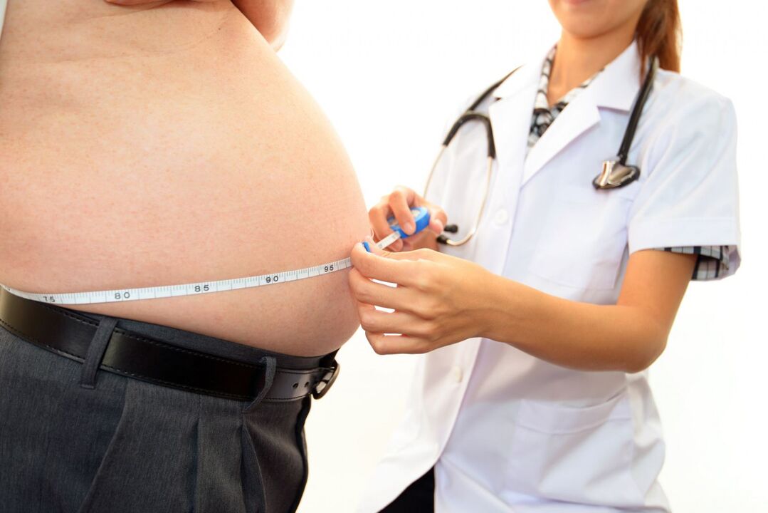
- Overweight.
- Endocrine system dysfunction (diabetes, psoriasis).
- Congenital pathology of musculoskeletal structure and development.
- Professional characteristics of labor activity.
- Poor local circulation.
- Previous diseases caused by pathogenic flora.
- Legg-Calve-Perthes disease.
- Metabolic disorders (gout).
- Physical inactivity.
- Immune diseases.
These reasons may not always cause ATS. Often, the activation of the pathological process can be provoked by:
- increased stress and physical activity;
- continuous overwork;
- vehicle or body hypothermia as a whole;
- lifting heavy objects suddenly;
- hormonal imbalance;
- radiation exposure.
Disease symptoms
Manifestations of ATS symptoms are similar to other joint arthrosis manifestations.
The main characteristic symptoms of this disease are considered to be:
- Stiffness in the morning or after long periods of immobility.
- Reduced range of motion, changes in gait.
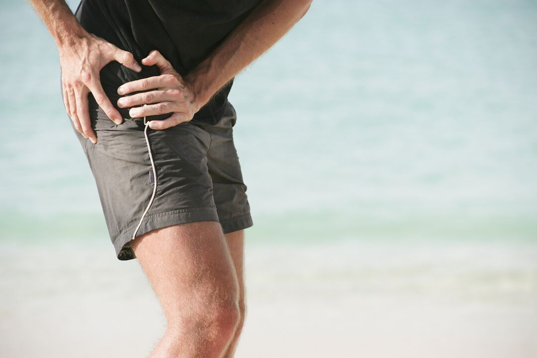
- Pain, first caused by mechanical or physical stress, then persistent.
- Manifestations of creaking, throbbing and clicking during sudden movements.
- Significant deformity of the affected limb.
- Occurrence of contractures (restriction of passive movement).
- Narrowing or closing of the joint space (X-ray sign).
The severity of the symptoms of arthrosis of the hip joint depends on the level of development of the disease and the reactive abilities of the patient's body.
Stage of coxarthrosis
Depending on the clinical manifestations, 4 stages of hip joint arthrosis can be distinguished:
- Arthrosis stage 1 of the hip joint does not have significant pain and other manifestations. A difficult stage to diagnose, this disease can be detected using the biochemical study of hyaline cartilage tissue and the determination of an insufficient amount of glycosaminoglycans. Patients feel pain in the joints and rarely pain at the beginning of physical activity.
- Arthrosis of the second degree of the hip joint is characterized by changes in the density and elasticity of cartilage fibers. Cracks and breaks appear. The depreciation function is reduced. The pain increases, radiating to the inguinal area, the movement of melting and reduction of the affected limb is limited.
- In the third stage, the stratification of cartilage fibers occurs with greater intensity. The articular surface is subjected to excessive pressure, the focus of ischemia develops. Cartilage grows along the edge of the epiphysis. The sensation of pain in the area of the damaged bone junction does not depend on the state of activity and rest. With any movement, the joint "creaks" and "beats". The range of motion is reduced on all axes.
- The fourth stage is characterized by exposure of the surface of the articular components with the formation of ulcers and depression. The articular head of the femur is not properly seated in the acetabulum, which leads to a comparative violation and separation of the articular surface. During this period, the patient experiences excruciating pain caused by narrowing, sometimes closing the articular lumen and compression of bundles of nerve fibers and blood vessels. Movement is limited, sometimes completely.
The classification of pathological changes caused by ATS is necessary to understand the mechanisms and characteristics of disease progression. Determining the severity of the disease helps determine the correct treatment tactics and disability (in case of severe disease).
Possible consequences
The progression of ATS leads not only to the deformation of the femoral head and pelvic cavity, but also to the development of pathological processes in the functioning of the articular apparatus as a whole.
Pathology due to hip arthrosis complications:
- synovitis (inflammation of the synovial membrane of the joint);
- aseptic necrosis of the femoral head;
- joint destruction (osteonecrosis);
- inflammation of the joint bag with changes in the amount of synovial fluid;
- partial or complete ankylosis (immobility of bone articulation);
- contracture (limitation of mobility and impossibility of flexion-extension of limbs).
The development of ATS complications always leads to a deterioration in the patient's general condition, quality of life and loss of unaided movement.
Diagnostic methods
Diagnosis of hip joint arthrosis in the early stages is difficult. Manifestations of symptoms become noticeable only when the bone epiphysis and nerve fibers are involved in the pathological process.
During the medical examination at the developmental stage, the following are observed:
- visual changes in the articular contour;
- palpable pain;
- sometimes pastosity of periarticular tissue;
- shorten the diseased limb.
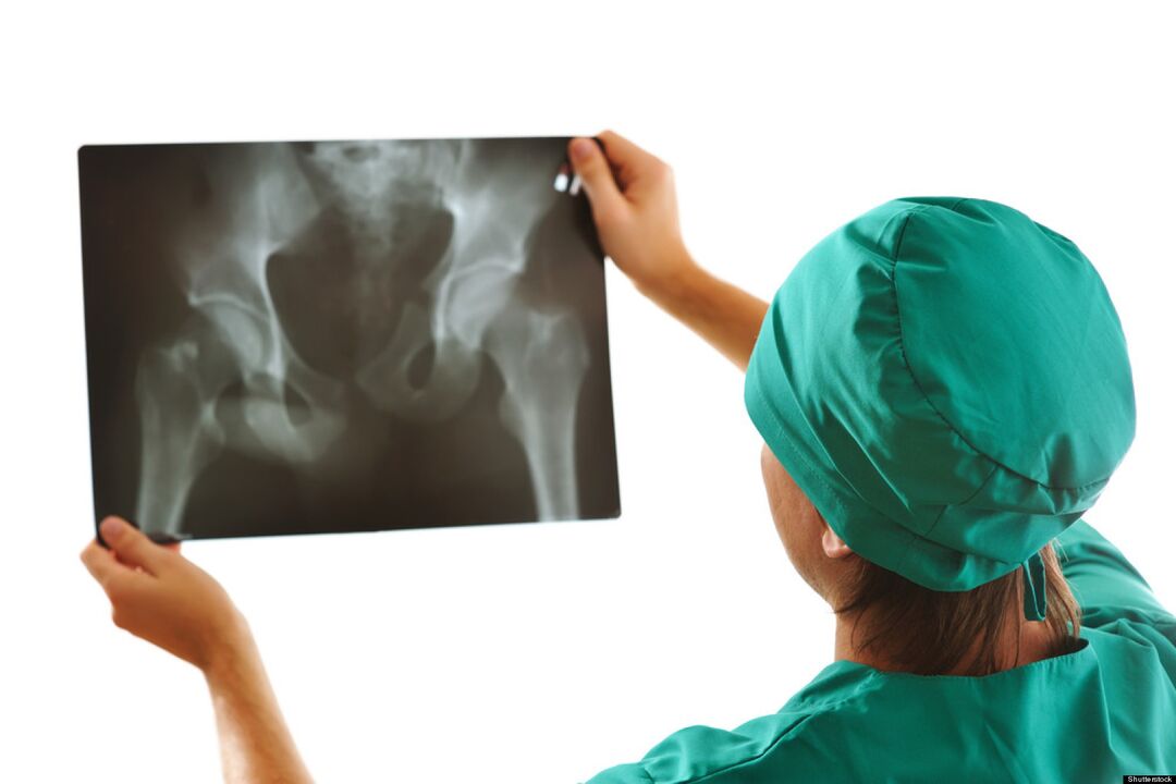
The main role in the diagnosis of ATS is given to X-ray examination. As an additional diagnostic method used:
- Ultrasound, magnetic resonance imaging.
- CT scan.
- Puncture of articular lubrication (synovial fluid).
- Diagnosis using an arthroscope (microscope).
- Clinical and biochemical laboratory tests of urine, blood.
Timely diagnosis improves the prognosis of treatment and further life of the patient.
How to apply for disability?
It is impossible to cure this disease completely. To confirm the right to social benefits and assign a disability group after passing an examination by a narrow specialist, you must contact your doctor.

Indications for the assignment of defects in the case of arthrosis of the hip joint are:
- oligoarthrosis (lesions of no more than 2 joints) TS 2 degree;
- combination of 2nd degree TS arthrosis and 3rd degree arthrosis of the knee joint;
- reduction in the length of the diseased limb by more than 6 cm;
- automatic phone exchange flows reactive, documented.
In determining the disabled group will help:
- carefully collected anamnesis;
- conclusions of the medical advisory commission (MCC);
- results of diagnostic studies;
- passed the medical and social expert commission (MSEC).
If the expert commission's decision is negative, it can be appealed to the superior.
Prevention
Preventive measures are a simple way to avoid the development of this disease. Preventive measures include:
- Adherence to an active lifestyle.
- Weight indicator control.
- Optimization of nutrition and work and rest methods.
- Reduce mechanical and physical load.
- Treatment of diseases of viral and infectious etiology.
- Prevention and prevention of injuries at home and at work.
- Regular preventive checkups.
Conclusion
The answer to the frequently asked question: "Is it possible to cure arthrosis of the hip joint? " Experts give a negative answer. Destroyed cartilage cannot be completely restored, just as it is impossible to completely correct the deformation and destruction of the bones included in the joint. Do not ignore even small manifestations of hip arthrosis, this reduces the chances of preventing the further development of the disease.